Pain and swelling after an injury should gradually become easier to manage, not remain unchanged or interfere with normal use of the limb. You will learn what to do during the first 72 hours, which symptoms require urgent care, when to arrange an assessment, and how to test whether you are ready to resume normal activity.
Key takeaways
- Expect clear improvement within days for most mild sprains and strains.
- Seek urgent care for severe pain, deformity, numbness, or inability to bear weight.
- Return to activity only when movement, strength, and function have recovered.
- Persistent swelling needs assessment when it limits use or stops improving.
How long should pain and swelling take to improve?
Most mild sprains and strains show clear improvement over several days and often settle within about two weeks. More severe injuries can take several weeks or months, so judge recovery by its direction and by how well you can use the limb, not by a fixed promise of complete healing.
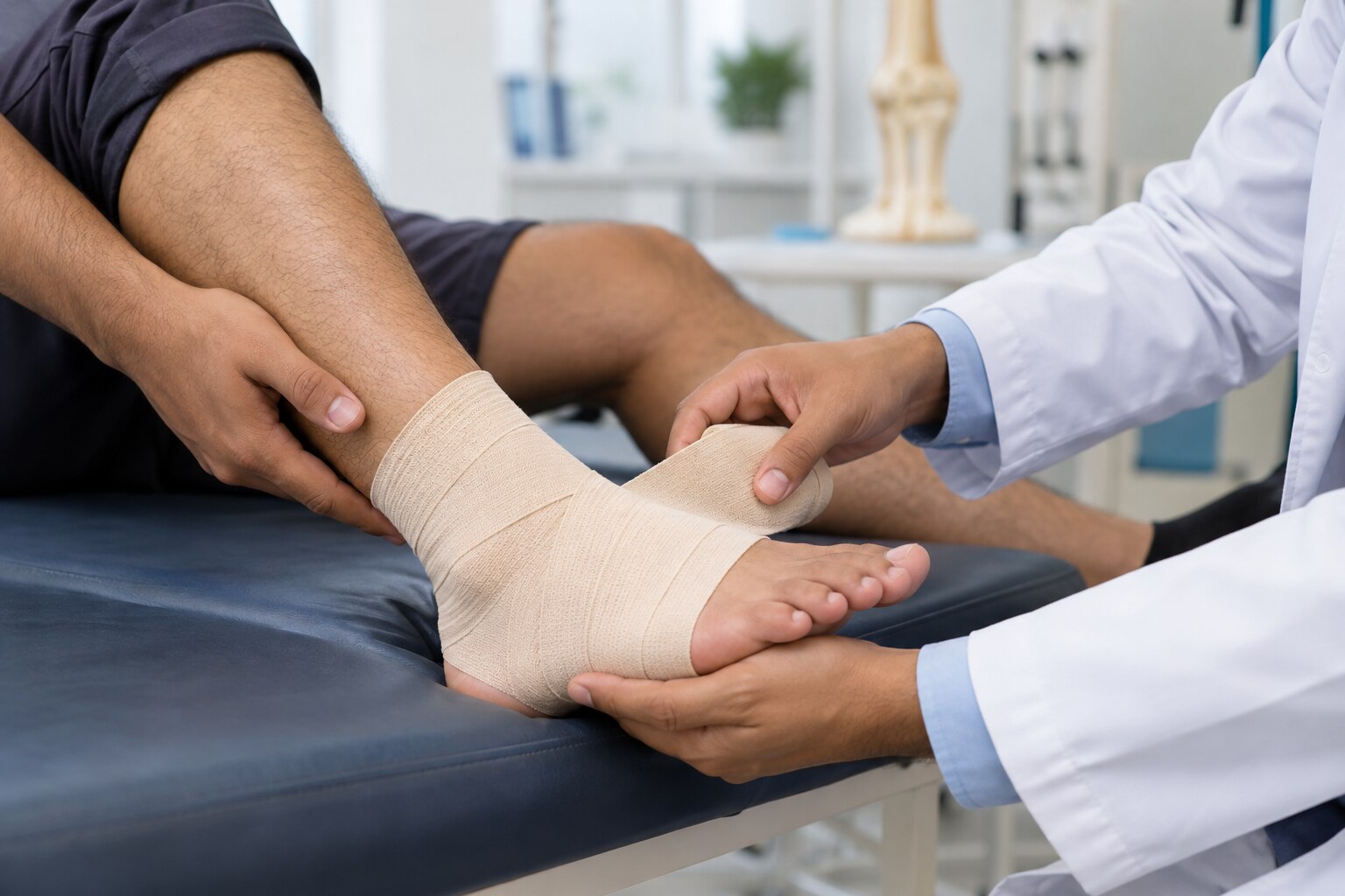
During the first 24 to 72 hours, stop the activity that caused pain and protect the limb from further strain. Use comfortable compression and elevate it above heart level when resting. This rest, ice, compression elevation approach can reduce discomfort and swelling.
Apply a cold pack wrapped in a cloth for short sessions; never put ice directly on skin. Do not sleep with a tight compression bandage or force painful movement. Skip ice over numb skin, broken skin, or an area with poor circulation. Ice is not enough when swelling is rapidly increasing or function is deteriorating.
Arrange an assessment if pain or swelling is not clearly improving after several days. Seek review sooner when substantial pain, bruising, swelling, or difficulty using the limb remains after one to two weeks. Persistent pain and swelling that limits walking, gripping, or normal joint movement needs attention rather than indefinite home care.
A clinician can check for a fracture, significant ligament or tendon injury, and the rehabilitation needed for safe recovery.
Which swelling and pain symptoms need urgent care?
Early swelling after a sprain is usually mild to moderate, appears within hours, and does not steadily worsen. Urgent care for a sprain is needed when pain or function suggests a fracture, dislocation, tendon rupture, nerve injury, or blood-vessel injury.
| Finding | More consistent with routine injury | Needs urgent assessment |
|---|---|---|
| Swelling and bruising | Mild and stable; movement remains possible | Severe bruising, rapidly increasing swelling, or inability to bear weight or use the limb normally |
| Shape and sensation | No deformity; normal colour and feeling | Visible deformity, numbness, tingling, blue or unusually pale skin, or loss of circulation |
| Sound and strength | Soreness without sudden weakness | An audible pop and weakness, which can indicate tendon or ligament rupture |
Pain out of proportion to the injury, marked tightness, or pain worsened by passive stretching are compartment syndrome warning signs. Acute compartment syndrome is an emergency; attend an emergency department immediately.
A hot, red, swollen joint with fever, chills, feeling very unwell, or rapidly worsening pain needs urgent assessment for infection. New calf swelling, warmth, redness, or tenderness after bed rest, immobilisation, or reduced mobility should not be labelled a muscle strain.
Chest pain, sudden breathlessness, coughing blood, or fainting requires emergency care because of possible deep-vein thrombosis and pulmonary embolism.
Seek assessment sooner after minor trauma if you are older, have diabetes, poor circulation, osteoporosis, immune suppression, or take anticoagulants. These conditions raise the risk of occult fracture, haematoma, infection, and delayed healing.
What should you check before returning to work, walking, or sport?
Reduced pain is not enough to prove recovery. Before return to activity after sprain, confirm pain-free range of motion, near-equal strength on both sides, and a weight-bearing test without limping.
1. Move the joint through its usual range. Stop if movement causes sharp pain, catching, or a blocked feeling.
2. Test strength against the uninjured side, then practise balance and proprioception exercises, such as standing on the injured leg. Include progressive range-of-motion, strengthening, balance, and proprioception work as treatment, not as an optional stage after symptoms disappear.
3. Progress in order: ordinary walking, stairs, normal work tasks, lifting, jogging, then sport-specific movements. If swelling increases later that day or the next morning, return to the previous level and arrange assessment if the setback persists.
4. Do not return to sport or heavy work until strength and balance are near equal. Faded bruising does not mean the tissues are ready; advancing too soon increases reinjury risk.
Recurrent giving-way, persistent instability, joint instability and locking, catching, inability to regain normal motion, or pain directly over a bone makes self-treatment the wrong choice. These symptoms can indicate ligament instability, cartilage injury, tendon damage, or an occult fracture and need clinical assessment.
Pain or swelling that prevents progression also warrants supervised rehabilitation rather than indefinite rest.
What happens during an assessment, and when are scans needed?
Assessment starts with the injury story: how the force occurred, whether you heard a pop, when swelling appeared, and whether you could walk afterward. The clinician locates the exact point of tenderness, checks swelling and bruising, and compares the injured limb with the other side.
The examination includes:
- Joint stability, range of motion, and strength
- Skin colour, temperature, pulses, and circulation
- Sensation, tingling, and other nerve function
- Ability to bear weight and take four steps
For an acute ankle injury, the Ottawa Ankle Rules support an X-ray for ankle injury when there is bone tenderness at the posterior edge or tip of either malleolus, or at the base of the fifth metatarsal or navicular.
An X-ray is also indicated when you cannot take four steps both immediately after the injury and during examination. These rules apply to selected ankle injuries, reduce unnecessary radiographs, and do not replace examination or apply automatically to every joint.
X-rays show many fractures and alignment problems, but not most ligament tears, tendon injuries, cartilage lesions, or bone-marrow injuries. For ankle pain lasting six weeks or longer, plain radiographs are generally the initial test.
MRI can assess ligaments, tendons, cartilage, marrow, and occult bone injury; ultrasound can assess selected tendons, ligaments, fluid collections, and movement in real time. A normal X-ray does not end evaluation when symptoms persist, so ask about ultrasound versus MRI based on the suspected structure.
When does persistent swelling require supervised treatment?
Persistent swelling needs supervised treatment when it prevents you from regaining motion, strength, balance, or normal walking. Most isolated ankle sprains improve without an operation through protection or bracing, early protected movement, progressive weight-bearing, and structured rehabilitation.
| Finding | Next step | What it may change |
|---|---|---|
| Mild swelling that is steadily decreasing | Continue graded home exercise | Keep progressing motion and weight-bearing |
| Pain, swelling, weakness, or limited motion that blocks progress | Arrange supervised rehabilitation | A clinician can adjust activity, choose a brace, and prescribe strengthening and balance work |
| Home exercises have not restored function | Seek an orthopaedic assessment for swelling | Examination may identify tendon, cartilage, fracture, or ligament problems |
| Repeated giving-way, catching, locking, or persistent ankle instability | Arrange prompt review and appropriate imaging | Mechanical instability or an occult injury needs a different plan |
A clinician can compare both ankles, test stability and strength, and decide whether an X-ray, MRI, or ultrasound is useful. A normal X-ray does not exclude most ligament tears, tendon injuries, cartilage lesions, or bone-marrow injury.
Surgery is reserved for selected cases, including persistent mechanical instability, certain fractures, or confirmed cartilage or tendon damage after proper evaluation and rehabilitation. A consultation with Dr. Santosh Pandey can match examination findings and imaging to a safe treatment plan when symptoms have outlasted the expected improvement window or normal activity remains restricted.
Seek assessment when recovery has stalled, function remains limited, or any red flag appears instead of repeating indefinite rest and ice.
Related service
Sports Injury & Medicine Sports Medicine Services focus on the prevention, diagnosis, treatment, and rehabilitation of sports-related injuries and musculoskeletal conditions... View service → |
Frequently asked questions
How long should sprain pain and swelling take to improve?
Most mild sprains and strains show clear improvement over several days and settle within about two weeks. Severe injuries can take several weeks or months.
Which sprain and strain symptoms need urgent care?
Seek urgent care for severe pain, visible deformity, numbness, loss of circulation, or inability to use or bear weight on the injured limb.
What should you check before returning to work, walking, or sport?
Check that pain and swelling are improving, movement is close to normal, strength has returned, and you can perform the required activity without worsening symptoms.
When are scans needed for a sprain or strain?
A clinician decides whether you need an X-ray, ultrasound, or MRI after examining the injury, especially when symptoms suggest a fracture, tendon injury, or persistent damage.
When does persistent swelling require supervised treatment?
Arrange an assessment when swelling stops improving, continues beyond the expected recovery period, limits movement or weight-bearing, or keeps returning with activity.