Knee pain that lasts for months can come from the joint itself, but it can also begin in the hip, spine, nerves, tendons or muscles. Knowing what records to bring, what movements and symptoms matter, and why a specialist chooses one test over another helps you prepare for a more useful evaluation and recognise symptoms that need urgent care.
Key takeaways
- Bring a symptom timeline, medication list, scan reports and prior treatment records.
- Describe triggers, swelling, locking, instability and pain location in specific terms.
- Urgent assessment is needed after major injury or with a hot, red, swollen knee and fever.
- Imaging and blood tests depend on examination findings, not pain severity alone.
What to Bring and How to Describe Your Symptoms
Bring a clear symptom timeline and every relevant record. Note which knee hurts, whether symptoms began after an injury or developed gradually, and how the problem has changed over weeks, months or years.
1. Write the pain location precisely: front, back, inner side, outer side, or behind the kneecap. Record pain onset, pain duration and severity, including whether it is constant, intermittent or worse at night.
2. Describe triggers and limits. Include walking distance, stairs, squatting, sitting, standing, turning in bed and rising from a chair. Say what relieves the pain.
3. Record swelling, warmth, clicking, catching, locking or giving way. State whether the knee ever becomes suddenly swollen and how long that swelling lasts.
4. List previous injuries, operations, injections and physiotherapy, including dates and how much each helped. Bring operative reports and physiotherapy or injection records if you have them.
5. Bring prior X-rays, MRI scans and their written reports, even if they seem old. Also take a medication list with doses, including painkillers, steroids and supplements.
6. Mention psoriasis, back pain, hip pain, numbness or tingling, fever, weight loss and swelling in other joints. These details can point away from a knee-only problem and help the specialist assess the hip, spine or nerves as well.
How the Examination Separates Knee Pain from Referred Pain
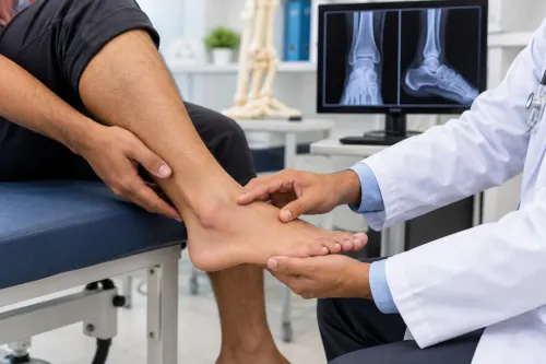
A knee examination tests whether the knee itself reproduces your pain, then checks nearby structures that can send pain into it. The specialist observes gait and alignment, including a limp, bow-legged or knock-kneed position, and quadriceps wasting.
The examination checklist includes:
- Range of motion: restricted bending or straightening, painful movement, and a fixed flexion contracture.
- Knee comparison: differences in warmth, shape, muscle bulk, movement and stability between the painful and unaffected sides.
- Tenderness and swelling: pain along the joint line, kneecap, patellar tendon, hamstring tendons or ligament attachments; an effusion suggests fluid inside the joint.
- Patellar tracking and meniscal signs: kneecap movement, clicking, catching, and pain during tests such as McMurray or Thessaly.
- Ligament stability: front-to-back and side-to-side movement using Lachman, drawer and collateral-ligament tests.
The pattern matters more than one manoeuvre. Local pain during resisted muscle or tendon loading points toward tendinopathy; excess movement after a twist suggests ligament injury. Joint-line tenderness with catching can support a meniscal problem, but no single test proves the diagnosis.
Hip rotation and hip range of motion are checked because hip arthritis can be felt near the knee. The specialist may examine the lumbar spine, leg strength, reflexes, sensation and nerve tension: back-related nerve irritation can cause burning, tingling or weakness around the knee without knee swelling.
Pain that changes with sitting, coughing or back movement strengthens that possibility.
Which Movement Findings Matter and Which Symptoms Are Urgent
An inability to fully straighten the knee can explain persistent discomfort, altered walking and difficulty standing from a chair. Reduced flexion limits squatting and sitting, while painful stair climbing often exposes load-sensitive joint, kneecap or muscle problems. Quadriceps weakness and wasting reduce shock absorption and can worsen instability, fatigue and pain.
| Finding | What it can explain | What it changes |
|---|---|---|
| Inability to fully straighten the knee | Limping, stiffness and reduced walking tolerance | Check for swelling, arthritis, meniscal blockage or another mechanical restriction |
| Reduced flexion | Difficulty squatting, kneeling or using stairs | Compare both knees and measure the actual range |
| Quadriceps weakness and wasting | Giving way, poor stair control and slower recovery | Assess strength and address it with suitable rehabilitation |
Seek urgent assessment for a suddenly hot, swollen and severely painful knee, particularly with fever, marked illness or inability to bear weight. Septic arthritis requires prompt exclusion; joint aspiration for synovial-fluid analysis and culture is a key test. A normal blood test or no fever does not safely rule out infection.
Morning stiffness lasting longer than about 30 minutes, several swollen joints, psoriasis, inflammatory back pain, unexplained weight loss or recurrent large effusions points away from simple wear-and-tear disease. Arrange timely specialist review, even if an X-ray shows only modest changes.
Pain and X-ray findings often disagree, so movement, daily function and treatment response matter more than an image alone.
When Examination Findings Lead to Imaging or Laboratory Tests
For chronic knee pain without a major recent injury, standing knee X-rays are usually the first imaging test. Weight-bearing views show joint-space narrowing, alignment and bone spurs under load; a skyline or sunrise view examines the patellofemoral joint.
X-ray changes do not measure pain: severe wear can cause little discomfort, while modest changes can be disabling.
| Finding | Investigation | What it can show |
|---|---|---|
| Persistent locking, giving-way or suspected internal injury with normal or limited X-ray findings | MRI for menisci and ligaments | Meniscal tears, ligament injury, cartilage damage, bone marrow changes and hidden joint disease |
| Focal tendon pain, suspected superficial fluid collection or an effusion | Ultrasound | Tendon thickening or tears, bursitis, cysts and fluid; it can guide an aspiration or injection but does not replace MRI for deep internal structures |
| Hot, swollen knee, fever, systemic illness or sudden severe pain | Blood tests and joint-fluid analysis | Blood tests such as complete blood count, C-reactive protein and erythrocyte sedimentation rate assess inflammatory or infectious clues. Aspiration tests white-cell count, Gram stain, culture and crystals |
| Sudden swelling without clear injury | Joint-fluid analysis | Monosodium urate supports gout and calcium-pyrophosphate supports pseudogout, but crystals do not exclude simultaneous infection; culture remains essential |
A normal blood test does not safely rule out septic arthritis. A suddenly hot, painful knee or inability to bear weight needs urgent assessment and aspiration rather than being labelled an osteoarthritis flare. MRI or ultrasound is chosen for the specific unanswered question, not simply because an X-ray looks abnormal.
How the Findings Shape Treatment and Further Decisions
Treatment follows the pattern of pain, lost function and objective findings, not an X-ray in isolation. A clinical osteoarthritis diagnosis can fit an adult aged 45 or older with activity-related pain and morning stiffness lasting no more than 30 minutes, even without imaging.
Structural damage and pain often disagree: severe X-ray changes can cause little pain, while modest changes can cause major disability.
- The specialist matches your symptoms with walking distance, stair use, sleep, work and examination findings. A small radiographic abnormality with severe instability, locking or weakness may prompt investigation for another problem rather than automatic arthritis treatment.
- The initial plan usually combines therapeutic exercise with activity adjustments and, when relevant, weight management. Losing weight is an adjunct, not a condition for receiving other care; a 10% loss generally offers more benefit than 5%.
- If pain blocks exercise, corticosteroid injections can provide short-term relief, roughly 2 to 10 weeks. They do not restore cartilage, so the response is judged by whether you can move and exercise more effectively, not by temporary pain reduction alone.
- Persistent symptoms can lead to review of the diagnosis, physiotherapy or another specialist referral. Dr. Santosh Pandey can use the treatment response alongside examination findings to decide whether knee-preserving care remains sensible.
- Knee replacement enters the discussion when pain and functional loss remain substantial despite appropriate non-surgical care, examination supports joint damage, and your goals align with the operation’s recovery and risks.
Related services
Physical Examinations ## Physical Examinations Physical examinations are a fundamental part of orthopaedic care, helping specialists assess your overall musculoskeletal... View service → |
Orthopaedic consultations ### Orthopaedic Consultations Orthopaedic consultations are the first step toward diagnosing and treating conditions affecting the bones, joints,... View service → |
Frequently asked questions
What should you bring to a chronic knee pain appointment?
Bring a symptom timeline, medication list, prior scan reports, treatment records and details of injuries or operations.
How does a knee specialist check whether pain comes from somewhere else?
The examination compares the knee with the hip, lower back and nerve function, then checks whether knee movements reproduce your symptoms.
Which knee symptoms need urgent medical assessment?
Seek urgent assessment for a hot, red, rapidly swollen knee, fever, inability to bear weight, severe injury or a locked knee.
When are imaging or laboratory tests used for knee pain?
The specialist chooses tests after examining your movement, stability, swelling and pain pattern; X-rays, MRI scans or blood tests answer different clinical questions.